Equine Gastric Disease The ins and outs (2024)
Don’t miss out on this essential information for every horse owner!
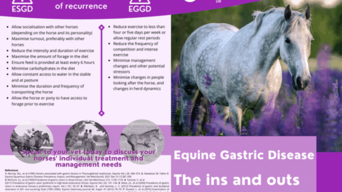
Reducing the risks of recurrence
- Allow socialisation with other horses (depending on the horse and its personality)
- Maximise turnout, preferably with other horses
- Reduce the intensity and duration of exercise
- Maximise the amount of forage in the diet
- Ensure feed is provided at least every 6 hours
- Minimise carbohydrates in the diet
- Allow constant access to water in the stable and at pasture
- Minimise the duration and frequency of transporting the horse
- Allow the horse or pony to have access to forage prior to exercise
- Reduce exercise to less than four or five days per week or allow regular rest periods
- Reduce the frequency of competition and intense exercise
- Minimise management changes and other potential stressors
- Minimise changes in people looking after the horse, and changes in herd dynamics
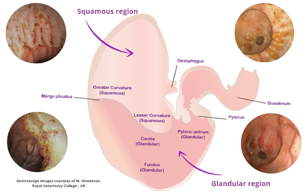
Equine gastric disease is the umbrella term for two disease types
Equine Squamous Gastric Disease (ESGD)
Occurs in the upper part of the stomach
The upper part of the stomach has little protection from acid exposure
Risk factors: High starch and sugar in the diet; Low forage; Intermittent feeding; Stress; High Intensity and duration of exercise
Mainly seen in horses eating high concentrate/low forage diets and exercising at high intensities
Equine Glandular Gastric Disease (EGGD)
Occurs in the lower part of the stomach
The glandular mucosa has protective mechanisms to prevent damage
Risk factors: High-frequency exercise with low rest periods; High-frequency competition and intense exercise; Lack of consistency with handlers and field friends
Seen in a wide range of horses, even those with more sedate lifestyles
Signs to look out for
Poor performance; Wind sucking; Lying down; Colic; Dull coat; Change in temperament to be handled; Change in ridden behaviour; Poor body condition; Reduced appetite; Girthy
The definitive method to diagnose equine gastric disease is via a gastroscopic examination
This procedure will take about 15-20 minutes.
Your horse will be lightly sedated. A three-meter long tube, with a camera on the end, known as a gastroscope, will be passed into the stomach via the horse’s nostril.
Your vet will examine all angles of the stomach from the squamous region, down into the glandular region.
Your vet will then discuss with you what has been seen, and if necessary a treatment and management plan.
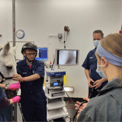
Image courtesy of M. Hewetson, Royal Veterinary College,UK
References
1. Murray, M.J., et al (1996) Factors associated with gastric lesions in Thoroughbred racehorses. Equine Vet J 28, 368–374. 2. Hewetson M, Tallon R. Equine Squamous Gastric Disease: Prevalence, Impact, and Management. Vet Med (Auckl). 2021 Dec 31;12:381-399
3. McClure, S.J., et al (1999) Prevalence of gastric ulcers in show horses. J Am Vet Med Assoc 215, 1130–1133. 4. Tamzali, Y., et al
(2011) Prevalence of gastric ulcer syndrome in high-level endurance horses. Equine Vet J 43, 141–144. 5. Nieto, J.E., et al (2004) Prevalence of gastric ulcers in endurance horses-a preliminary report. Vet J 167, 33–37. 6. Elfenbein, R. and Sanchez, L. C. (2012) Prevalence of gastric and duodenal ulceration in 691 non-surviving foals (1995–2006). Equine Veterinary Journal 44, Suppl. 41 (2012) 76–79. 7. Husted, L., et al (2010) Examination of equine glandular stomach lesions for bacteria, including Helicobacter spp by fluorescence in situ hybridisation. BMC Microbiol. 10, 84. 8. Hepburn, R. and Proudman, C.J. (2014) Endoscopic examination of the squamous and glandular gastric mucosa in sport and leisure horses: 684 horses (Abstract). Proceedings of the 11th International Colic Symposium.


